
The Curious Case of the Limping Child: Diagnosing Köhler Disease Early
A pediatric limping case highlighting the importance of early imaging and careful X-ray interpretation in diagnosing Köhler Disease.

Case Presentation
A 4-year-old boy presented to the Emergency Department with a limp and pain in the left foot lasting for two days.
History
- Mother denied any history of trauma or falls.
Examination
- Mild swelling and tenderness were noted over the dorsum of the left foot.
- Vital signs were all within normal limits.
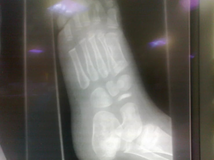
An X-ray of the affected foot was performed.
What Is the Diagnosis?
Upon careful review of the X-ray:
- The navicular bone appeared sclerotic, fragmented, hyperdense, small, and irregular.
This classic appearance leads to the diagnosis:
Köhler Disease
Understanding Köhler Disease
What is Köhler Disease?
- A rare idiopathic condition caused by avascular necrosis of the navicular bone.
- Results in painful limp and localized tenderness over the navicular area.
Historical Note
- First described by Dr. Alban Köhler, a German radiologist, in 1908.
Etiology
- May follow minor trauma or occur without any prior injury.
Classification
- Belongs to the group of crushing osteochondritides.
Epidemiology
- Prevalence: 2% of children.
- More common in boys (male to female ratio 5:1).
- Affects children aged 3–7 years.
- Usually unilateral, but bilateral involvement occurs in 25% of cases.
Radiographic Features
In Köhler Disease, the navicular bone typically appears:
- Sclerotic and hyperdense
- Fragmented and small
- Irregular compared to the normal navicular bone
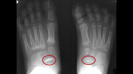
A comparative image may be shown highlighting the difference between normal and necrotic navicular bones.
Treatment
Conservative Management
- Pain control with rest and NSAIDs.
- Immobilization using a plaster cast (POP) in cases with severe symptoms.
- Orthopaedic follow-up is recommended.
Prognosis
- Excellent prognosis.
- Most cases resolve spontaneously within 6 to 48 months.
- No long-term disability reported.
Further Learning
References
Key Takeaways
- Always consider Köhler Disease in young children with an unexplained limp and localized foot pain.
- Early imaging is crucial to avoid misdiagnosis.
- Conservative management leads to full recovery without long-term complications.
Stay tuned for more intriguing pediatric radiology cases!
Want More Clinical Cases Like This?
Explore more practical case-based learning in our Emergency Radiology Cases section and continue building your clinical pattern recognition.
Frequently Asked Questions
What is Köhler Disease?
Köhler Disease is avascular necrosis of the navicular bone in children, leading to pain and limping, often resolving spontaneously with conservative treatment.
What are the common radiographic features of Köhler Disease?
The navicular bone appears sclerotic, hyperdense, fragmented, small, and irregular on X-rays.
How is Köhler Disease treated?
Treatment is conservative, involving pain management, rest, possible immobilization in a cast, and follow-up with orthopaedics.